
|
||
| Menu | OPAL / Imaging | |
|
OPAL Home About People International Collaborators
Opportunities
Events
Related Links
|
Imaging of the OPAL complexAcquiring a diverse set of medical data provides subject-speci fic insight into the morphology of the OPAL structures as well as its dynamics during speech, chewing and swallowing. Static imaging techniques, such as: X-rays, Computed Tomography (CT) and Magnetic Resonance Imaging (MRI), capture the tongue in a sustained posture. The common issues to be addressed include the motion artifact caused by the relatively-long scan times, the poor signal-to-noise ratio, the insufficient spatial resolution and the low soft-tissue contrast. In addition, the approved level of the ionization dosage is a matter of active discussion. Dynamic imaging techniques, such as dynamic MRI, Videofluoroscopy, CT and Ultrasound, compete to record the nature and the chronology of the tongue movements during swallowing. As the whole swallowing process is done in less than 1.5 seconds, scan times of more than 300ms per frame can cause severe motion artifacts. Videofluoroscopy for diagnosis of dysphagia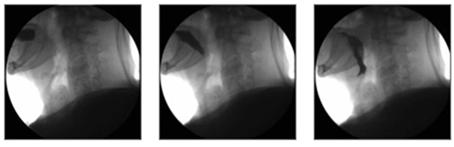 Videofluroscopyof the swallowing: capturing the bolus movement. Swallowing disorders, also known as dysphagia, can occur during oral, pharyngeal, esophageal phase of the swallowing process. General signs of dysphagia may include difficulty trying to swallow, coughing while swallowing, weak voice and weight loss. Dysphagia, in extreme cases, makes people totally unable to swallow and eating a challenge. Often people suffering from dysphagia cannot take in enough calories or fluids to nourish the body which leads to serious medical conditions. Video fluoroscopy (VFS) provides high spatial and temporal resolution and hence, has been regarded as the gold standard for evaluation of swallowing. The oropharyngeal region is exposed to X-ray radiation in upright or supine position, while the patient is swallowing Barium suspensions of different viscosities. The projection from the lateral view is then recorded in a video format. MBSImP (Modified Barium Swallow Impairment Profile)Modified Barium Swallow Impairment Profile (MBSImP) is a web based training tool to help clinicians gather more objective and standardized information on swallowing impairment. Bonnie Martin-Harris [http://academicdepartments.musc.edu/chp/directory/faculty/martinharris.htm] and her colleagues at the Medical University of South Carolina (MUSC) and Saint Joseph's Hospital of Atlanta, Georgia created MBSImP based on judgments of structural movement relative to bolus flow from videofluoroscopic images using standardized bolus volumes and consistencies. The MBSImP includes 17 physiologic components starting from lip closure to esophageal clearance. MBSImP gives a standardized protocol and provides a web-based learning platform to improve the accuracy and quality of MBS practice. This also gives the clinicians means to communicate MBS study results in a standardized, evidence based manner. More information can be found on the Northern Speech Services (NSS) website [http://www.northernspeech.com/MBSImP/]. Super resolution reconstruction of MRI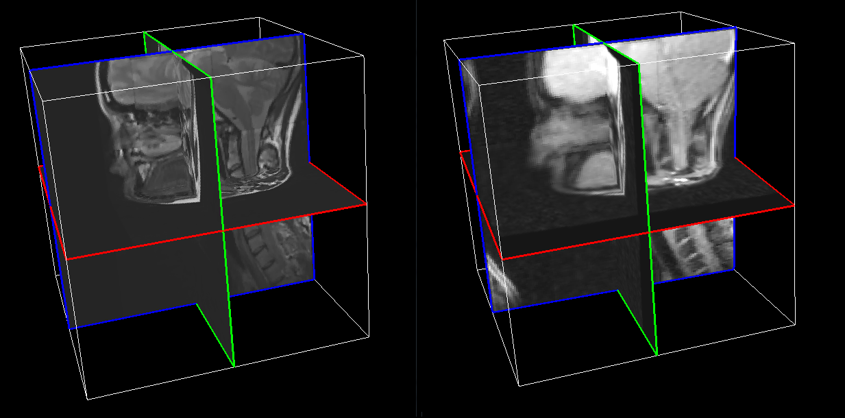 Super resolution reconstruction of static (left) and dynamic (right) MRI of the tongue. Detailed biology of the OPAL complex can be inspected with MRI. High-resolution MR volumes require a long acquisition time, which leads to involuntary movement of the tongue and, hence, introduces severe motion artifacts. 2D acquisition can provide a refined depiction of the tongue in the acquired plane, but the through-plane resolution is low, and inadequate for most of the volumetric analyses. Some previous speech studies adjusted the orientation of the acquisition plane to be orthogonal to the axis of the vocal tract, in order to facilitate the tongue modelling. Then, 2D tongue contours are manually segmented in each slice and registered to the volume. In a different approach, super-resolution reconstruction techniques have been introduced to generate isotropic MR volumes from orthogonal slice stacks acquired sequentially. First, the imaging process is formulated as an observation model. Next, the intensity value of each pixel in the volume is estimated, using an optimization method such as maximum a-posteriori (MAP) or least square(LS) iteratively. Recently, OPAL researchers, Stone et al. applied an edge-preserving data combination technique based on Markov Random Field, to build super-resolution volumes of the human tongue. Isotropic resolution of 0.94 mm was reported. Dynamic imaging can provide insight into the physiology of the tongue for different subjects. Stone et al., acquired dynamic tagged and cine MR image sequences, using consistent protocol, during the speech task. The images are acquired at multiple parallel axial slice locations covering the tongue. The resolution scheme is 1.875 mm in-plane (dense) and 6.00 mm through-plane (sparse). Super resolution MRI volumes are further reconstructed with an isotropic resolution of 1.875 mm, for each of the 26 time frames. Jonghye Woo, Maureen Stone and Jerry Prince (2012) Reconstruction of High Resolution Tongue Volumes from MRI.. . (BibTeX) Fangxu Xing, Jonghye Woo, Emi Z. Murano, Junghoon Lee, Maureen Stone and Jerry L. Prince (2013) 3D Tongue Motion from Tagged and Cine MR Images. In Proceedings of Medical Image Computing and Computer-Assisted Intervention(MICCAI)., pages 41-48, . Springer Berlin Heidelberg. (BibTeX) Dynamic CT of Swallowing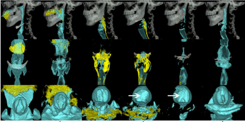 Dynamic CT of swallowing. Lateral (top row), AP (middle) and IS (bottom) views. Computed Tomography (CT) generates cross-sectional or volumetric images of high spatial resolution. CT is well suited for depiction of the contrast between the soft tissue and the air or bony structures. Multi-slice CT (MSCT) scanners have been recently equipped with multiple arrays of X-ray detectors. Our colaborators, Fujii et al., used a 320-detector-row MSCT scanner to capture a single phase 3D image volume of the oropharynx in less than 0.35 sec. The process was repeated for 29 phases at intervals of 0.1 sec to generate the full 3D video of the swallowing for one volunteer. The current state of the art allows for spatial resolutions of (0.5mm x 0.5mm x 0.5mm x 10 Hz). Fujii, Naoko, Inamoto, Yoko, Saitoh, Eiichi, Baba, Mikoto, Okada, Sumiko, Yoshioka, Satoshi, Nakai, Toshiaki, Ida, Yoshihiro, Katada, Kazuhiro and Palmer, JeffreyB. (2011) Evaluation of Swallowing Using 320-Detector-Row Multislice CT. Part I: Single- and Multiphase Volume Scanning for Three-dimensional Morphological and Kinematic Analysis.. . (URL) (BibTeX) Inamoto, Yoko, Fujii, Naoko, Saitoh, Eiichi, Baba, Mikoto, Okada, Sumiko, Katada, Kazuhiro, Ozeki, Yasunori, Kanamori, Daisuke and Palmer, JeffreyB. (2011) Evaluation of Swallowing Using 320-detector-row Multislice CT. Part II: Kinematic Analysis of Laryngeal Closure during Normal Swallowing.. . (URL) (BibTeX) Visible Korean Human Dataset (VKH)Image-Based 3D Modeling of the Complex "Tongue, Soft Palate & Pharyngeal Wall", <Dr. N. Amor> These high resolution anatomical images could be utilized to create any organ of the human body. Both soft (larynx, pharynx, soft palate, tongue, muscles, nerves, etc) & hard (maxilla, mandible, hyoid, vertebra, etc) tissues could be extremely distinguishable & consequently well-captured (2D, segmentation) & created (3D). The main focus of the ongoing research is to create a 3D model of the complete pharyngeal wall, tongue & soft palate system; a very delicate anatomical structure in the human body. To achieve this goal, one of the most valuable imaging software "Amira" developed by Visualization Sciences Group is being used. Images are imported into this software, followed by segmentation & registration of the concerned anatomical parts; essential steps for the generation & meshing of the corresponding 3D model. This will be implemented into Artisynth toolkit developed by OPAL team, and added to an existing (3D) model to complete the OPAL complex. The elaborated final 3D model could be used to simulate speech production, mastication &/or swallowing (Artisynth). Methodology |
|
| View Edit Attributes History Attach Print Search Page last modified on January 15, 2018, at 12:59 PM | ||